



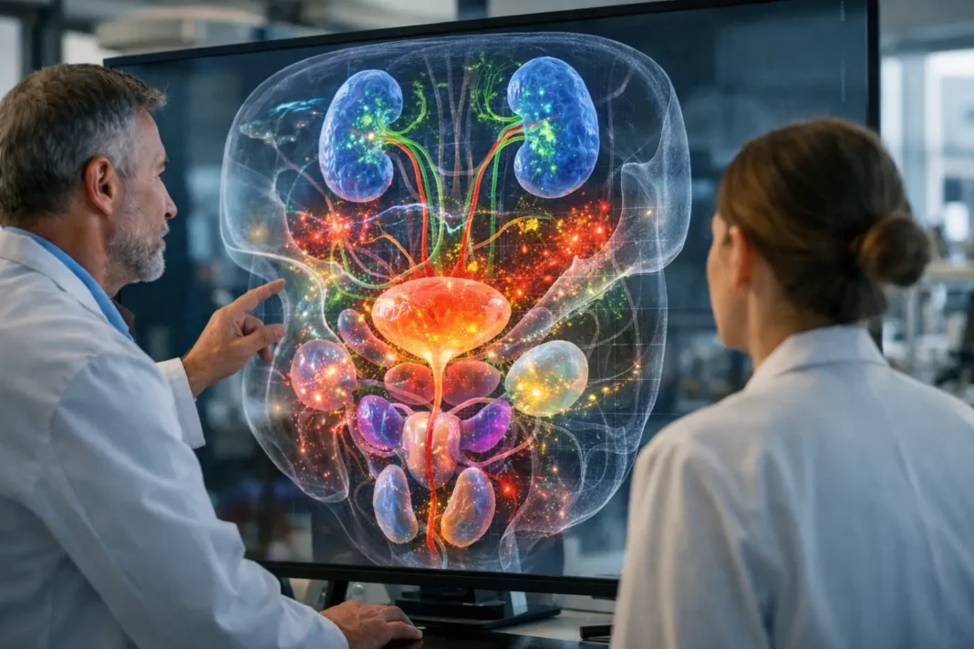
A new spatial map of embryonic development is offering a clearer view of how human urogenital organs form in relation to one another, rather than as isolated structures. The study follows organogenesis in its native spatial context, an approach that preserves the physical relationships between developing tissues. By doing so, it highlights how close anatomical neighbors exchange information while the urinary and reproductive systems take shape.
The research focuses on a crucial phase of human development, when early tissues begin to organize into distinct organs with specialized functions. In the urogenital region, this process is especially complex because multiple structures emerge in close proximity and must develop in a coordinated way. The mapping shows that cell-to-cell communication and molecular signaling are central to that coordination, helping explain how adjacent organs influence one another during formation.
For biomedicine, the value of such work lies in the level of detail it brings to an area that is difficult to study directly. Traditional developmental models can describe broad stages of organ formation, but spatial mapping adds another layer: where different cell types are located, how they interact, and how signals may travel across developing tissue boundaries. This makes it possible to interpret organ growth as a shared regional process rather than a sequence of separate events.
The findings also matter for research into congenital conditions affecting the urinary and reproductive systems. A more precise reference map of normal development can help scientists identify when and where developmental pathways diverge. While the study does not itself establish clinical applications, it strengthens the foundation for future work on disease mechanisms, regenerative medicine and laboratory models designed to reproduce human organ development more accurately.
Spatial biology has become an increasingly important tool in medical research because it combines molecular data with tissue architecture. In the case of human urogenital organogenesis, that combination helps clarify how structure and signaling evolve together. The study’s central contribution is therefore not only a map of developing organs, but a framework for understanding how neighboring tissues cooperate during one of the most intricate stages of human development.
